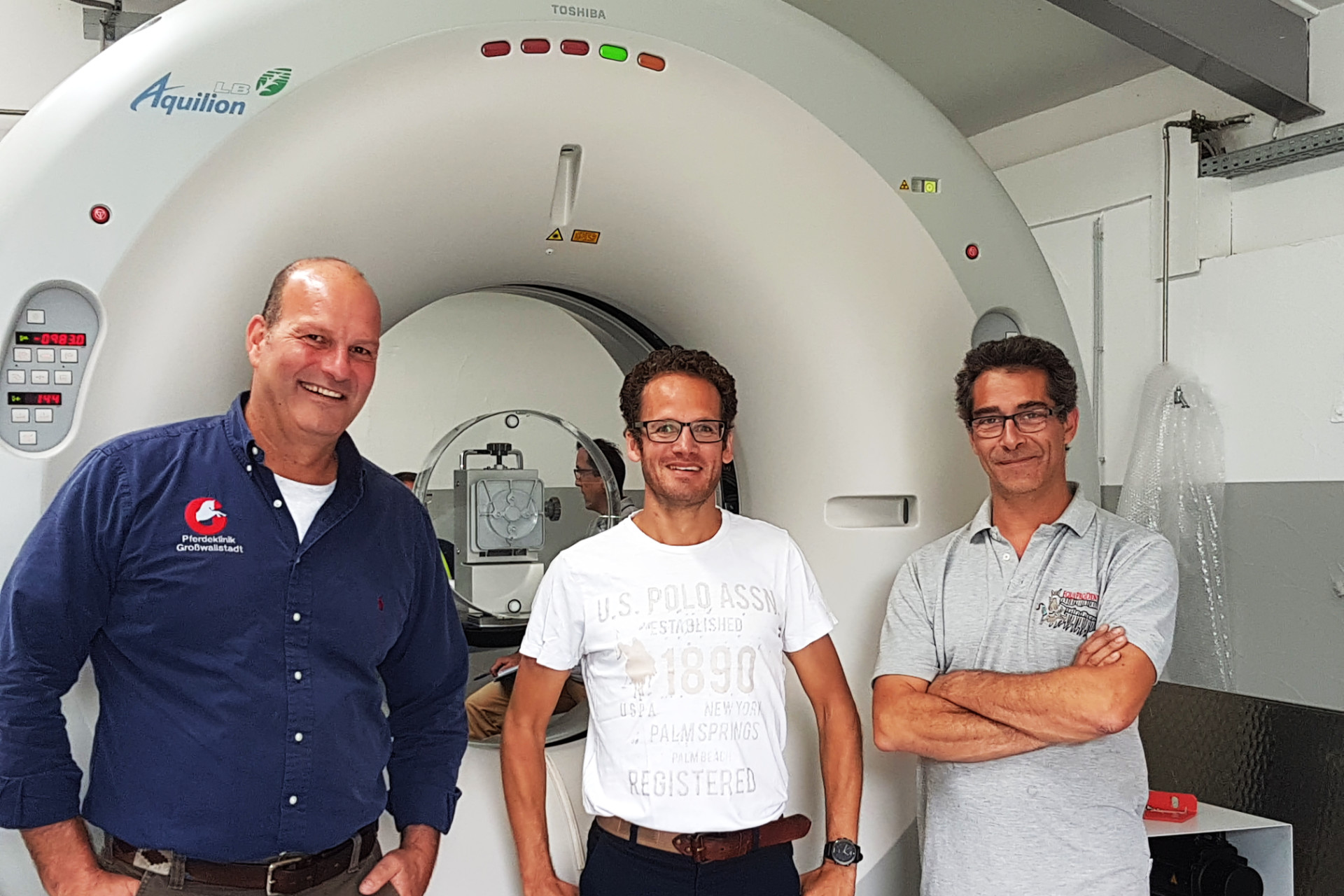
The team of the Veterinary Competence Center for Horses Großwallstadt Altano GmbH and the EquiProDenta are pleased to announce that from now on we also offer computed tomography (CT) examinations.
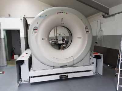
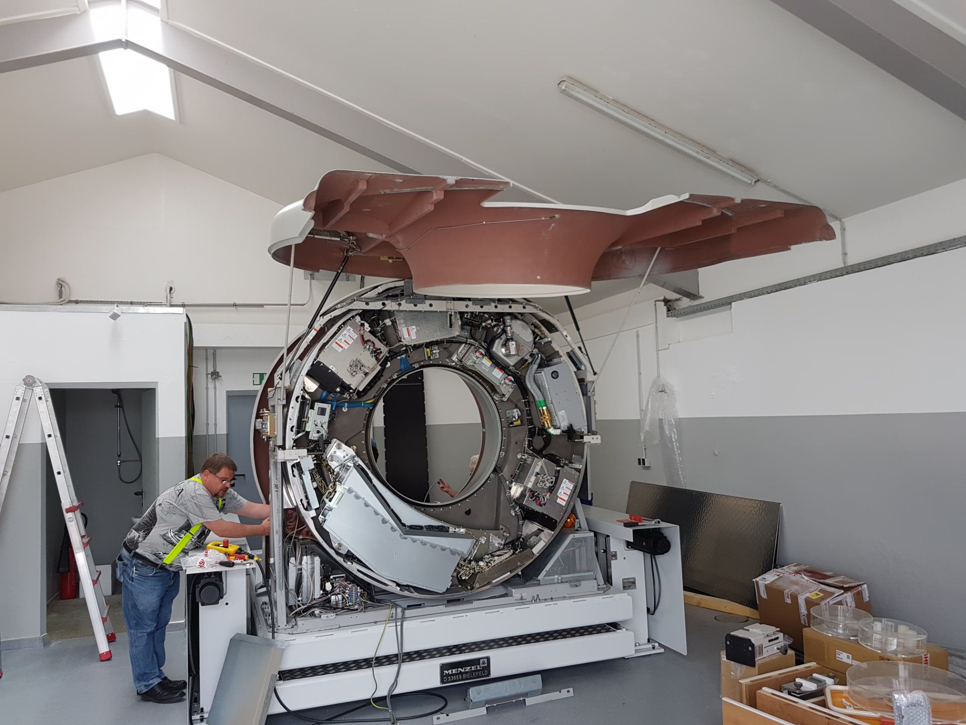
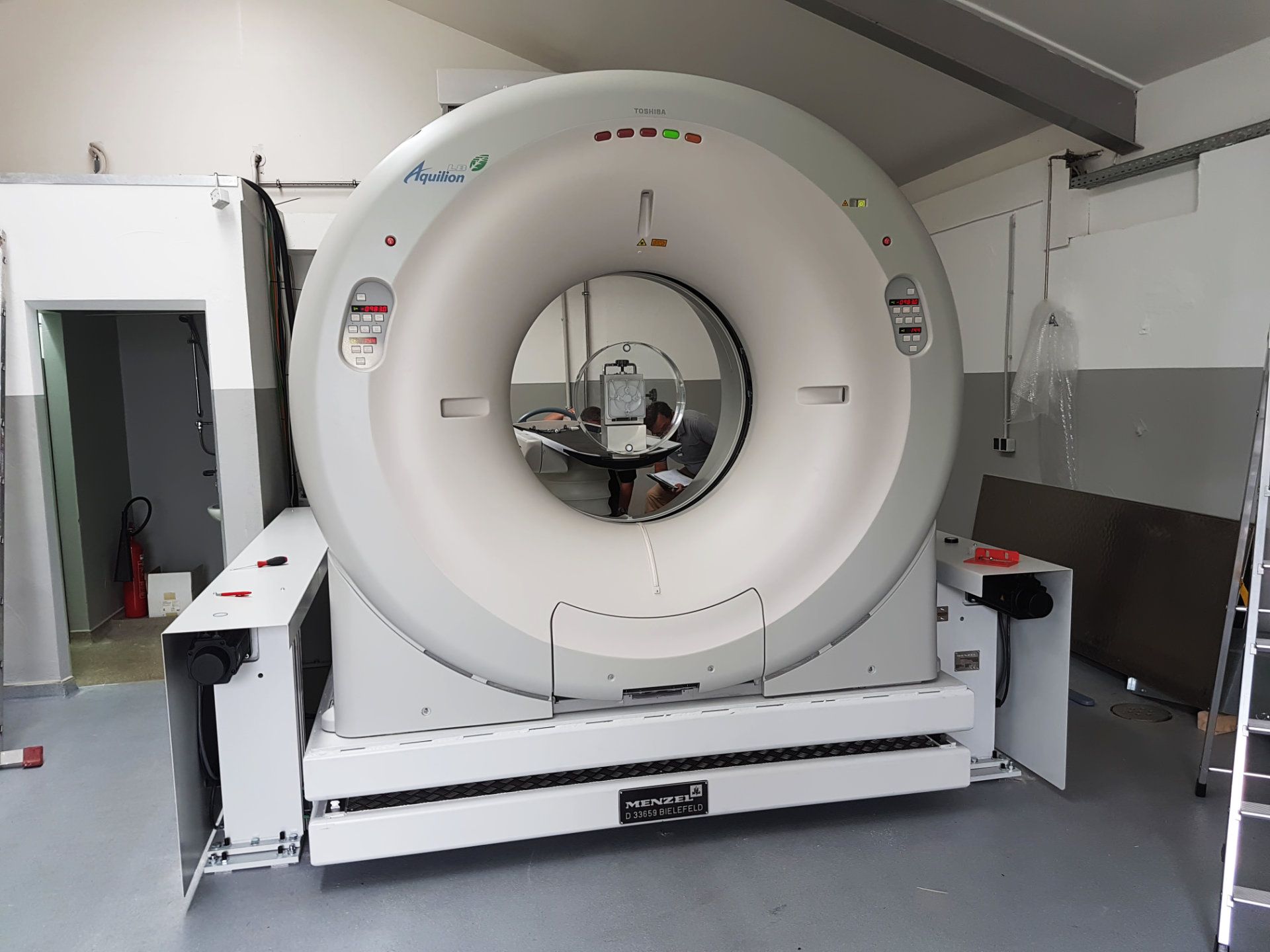
The Aquillion device from Toshiba has been strengthening the diagnostic capabilities of our clinic for a few weeks now and offers our equine patients an examination at the highest level.
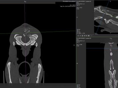
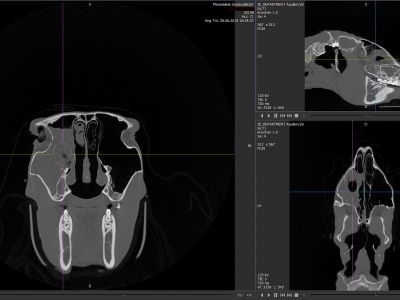
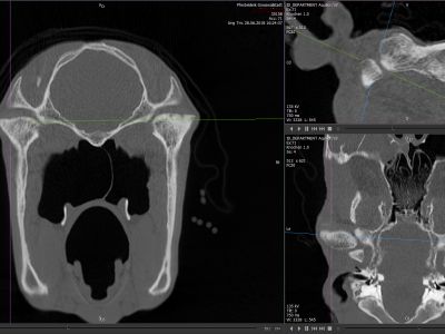
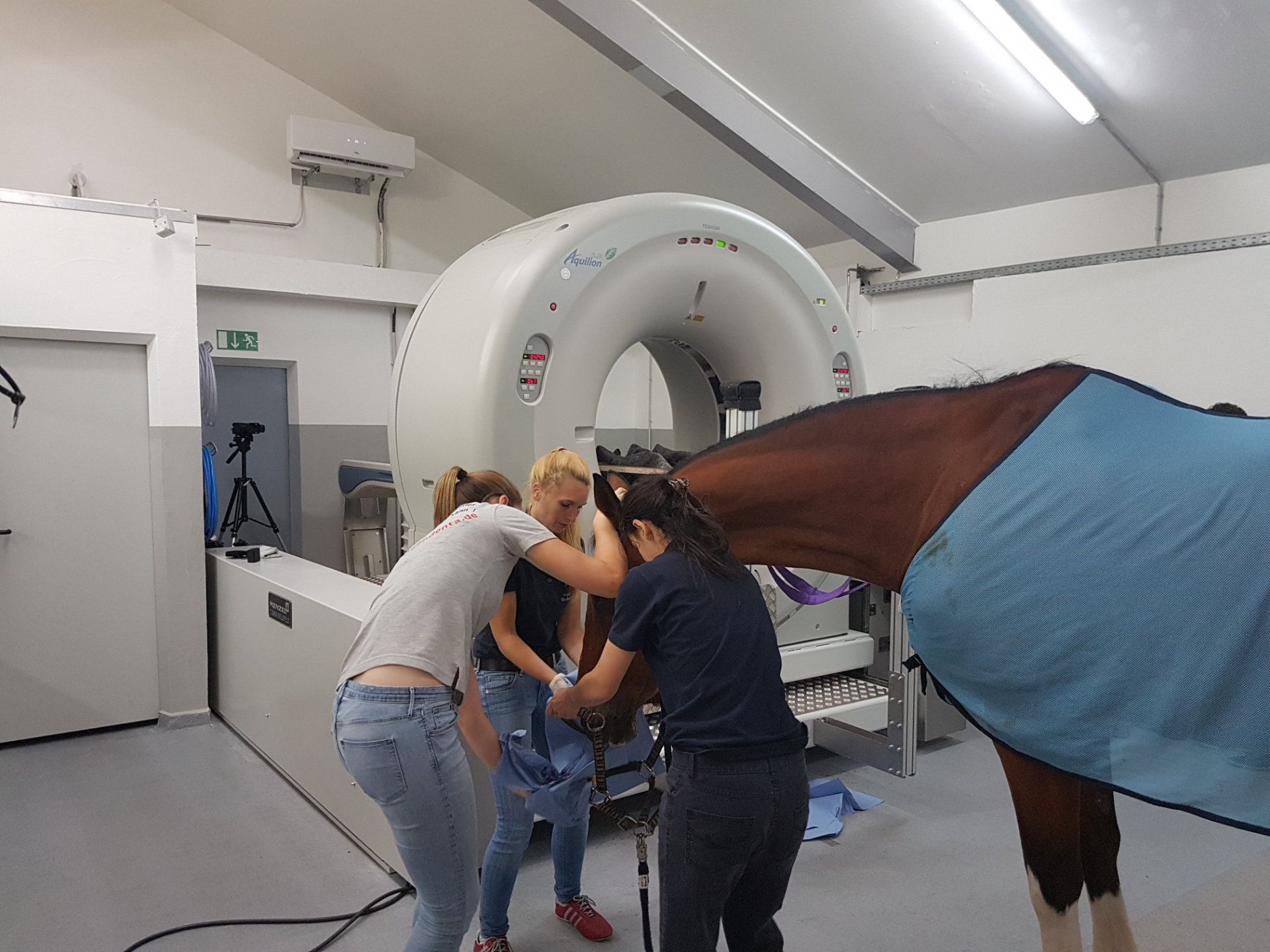
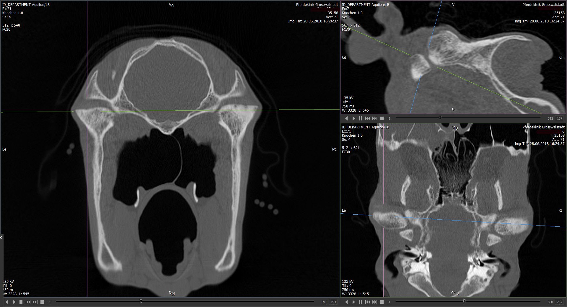
It is a 32-rower, this is considered the "gold standard" in veterinary medicine. Due to its large opening (gantry), the horses’ head and neck can be examined standing, and the distal limbs under general anesthesia, in less than 30 seconds.
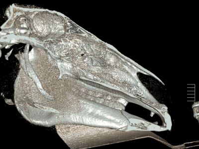
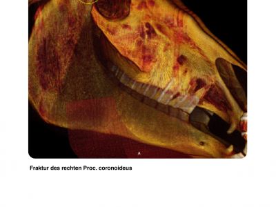
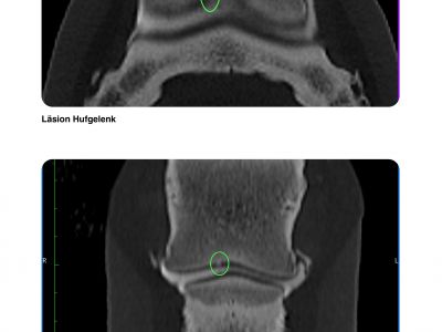
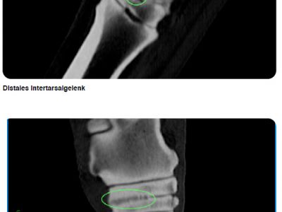
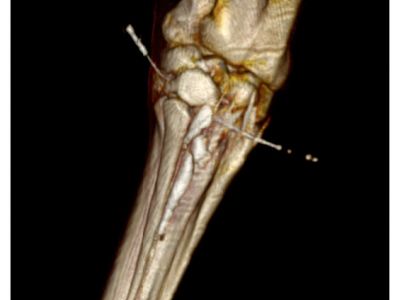
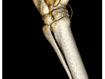
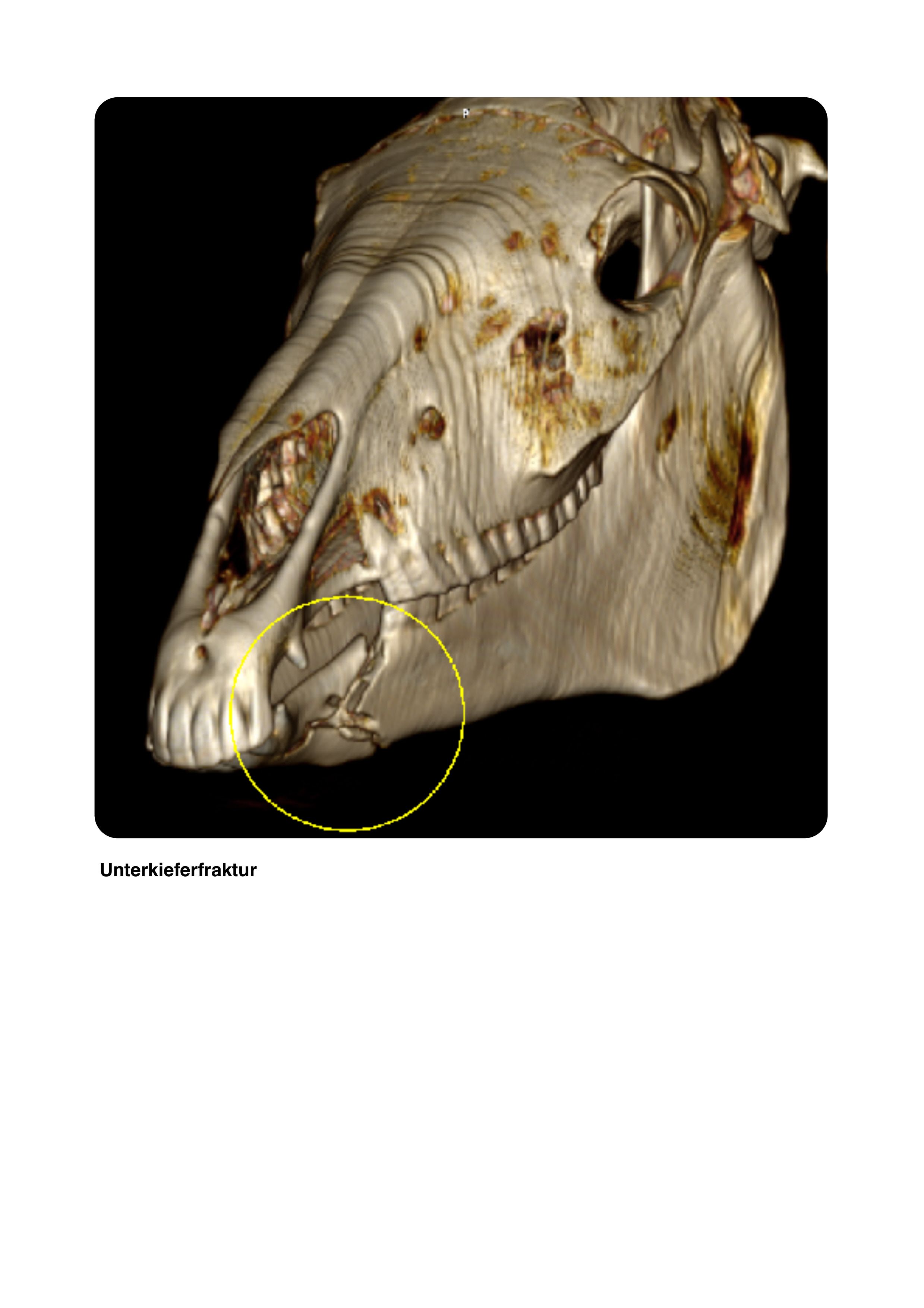
Computed tomography (CT) is an multi-dimensional X-ray examination. The generated slice images (0.5 mm thick) provide a detailed 3D view of the affected region of the horse's body.
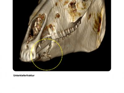
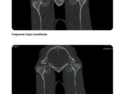
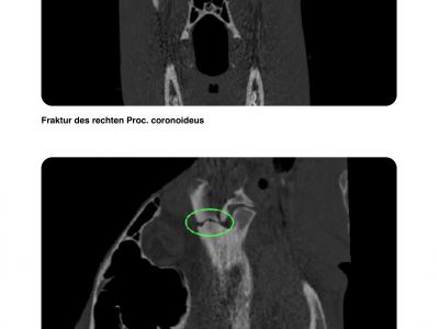
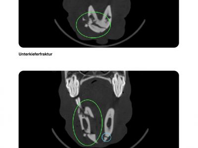
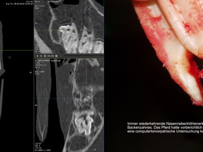
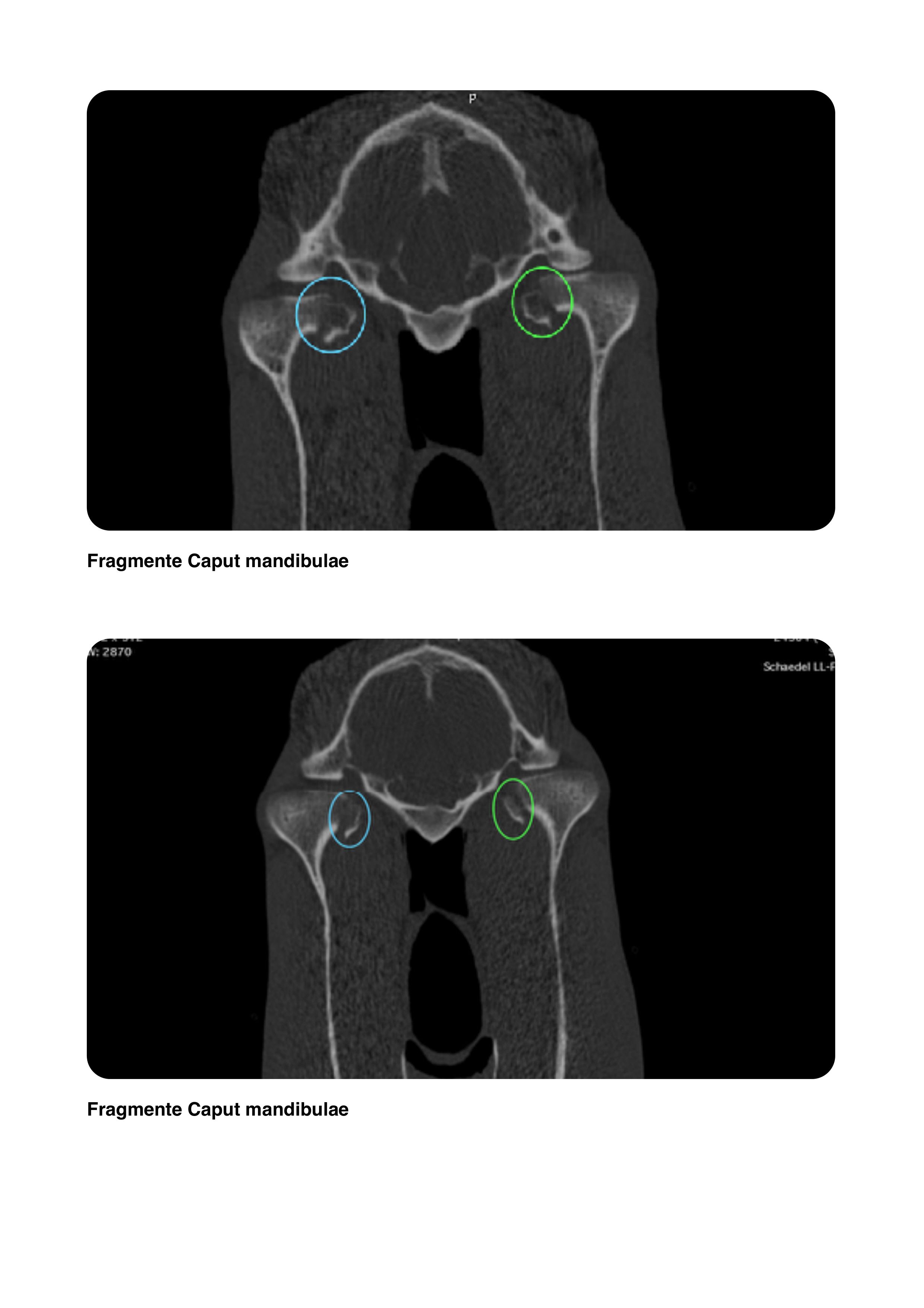
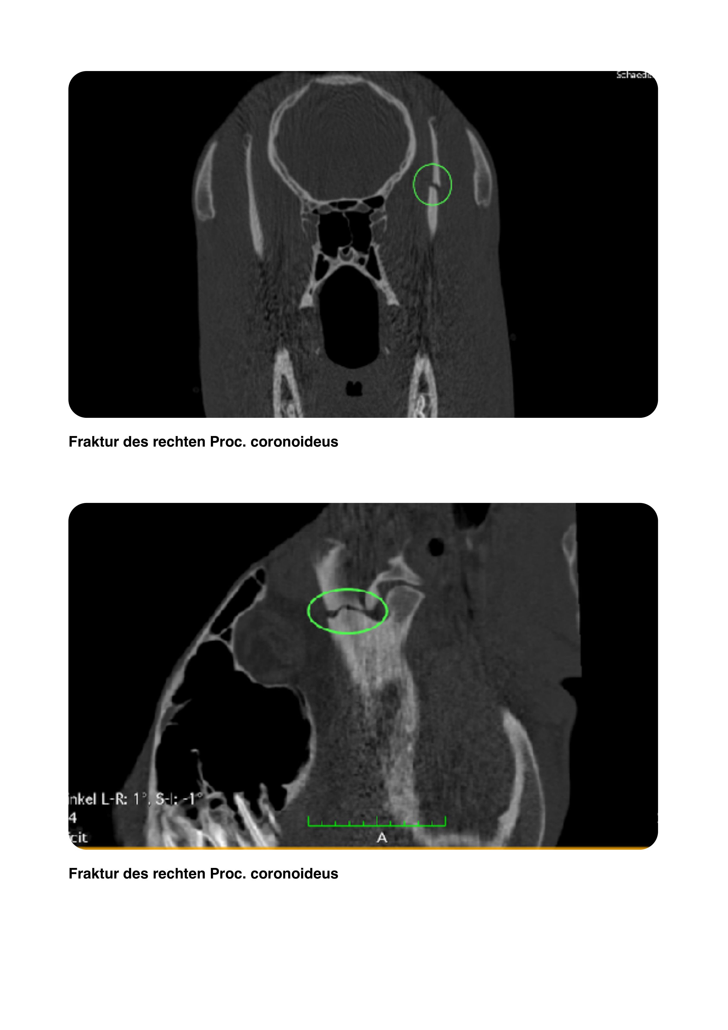
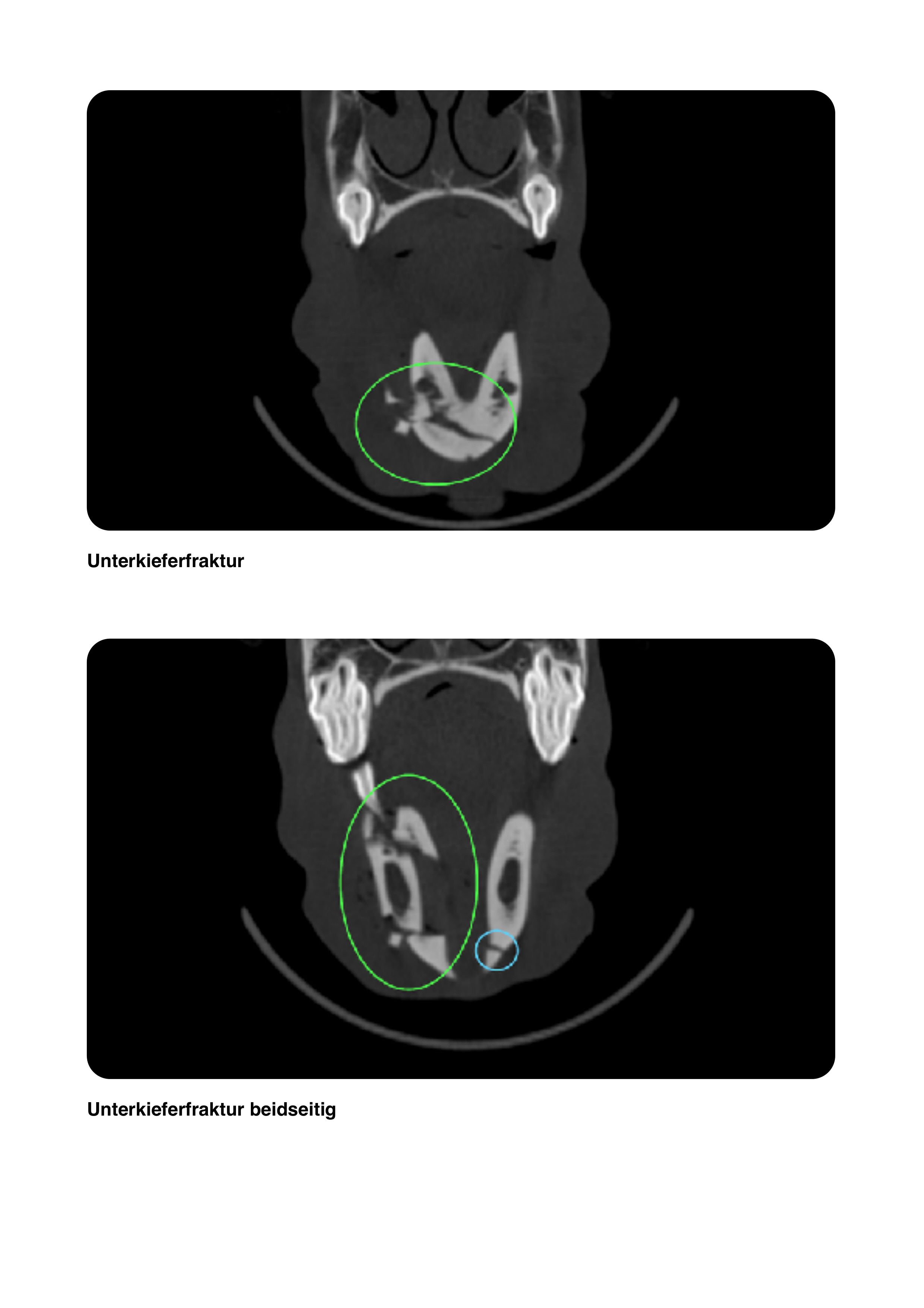
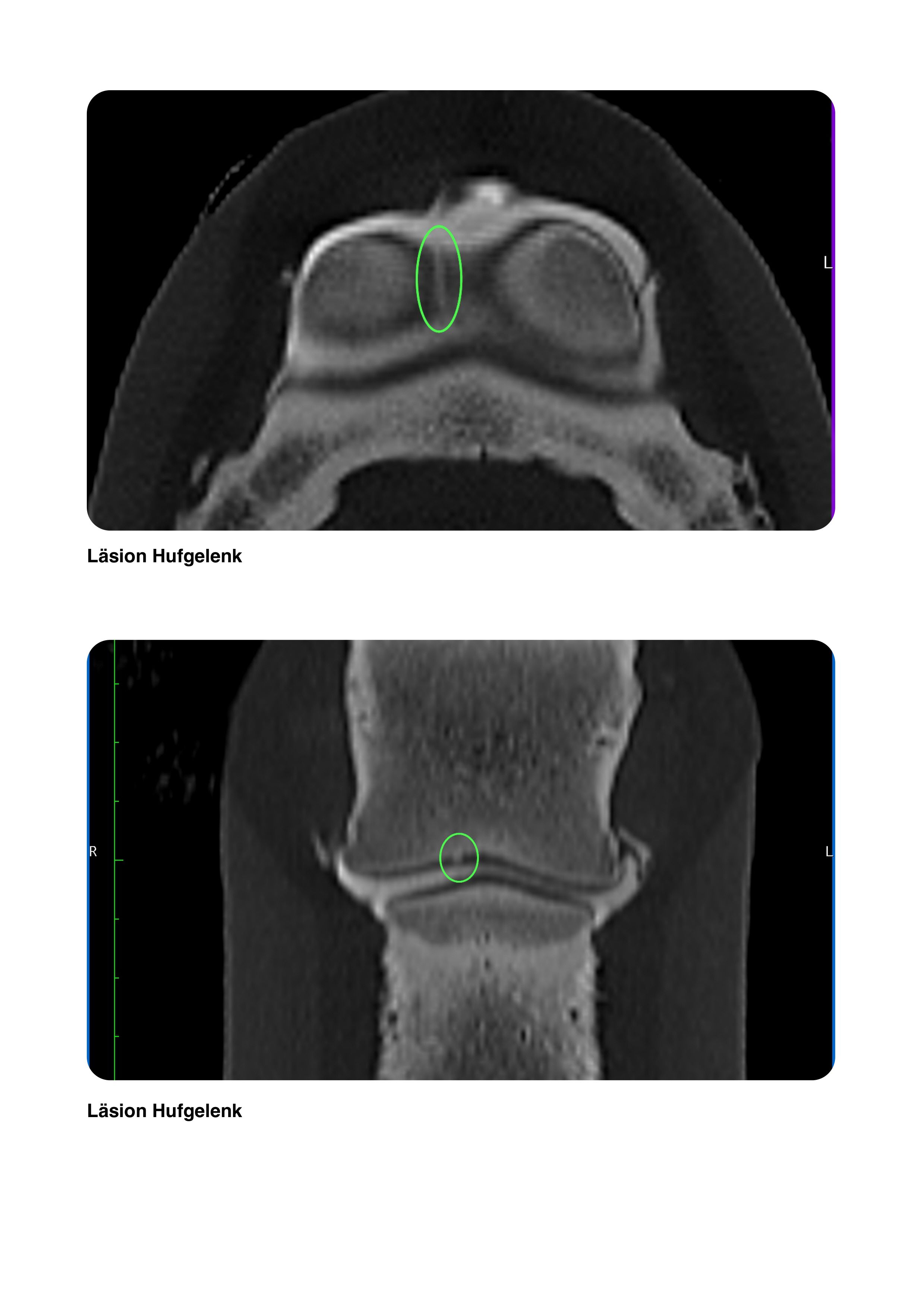
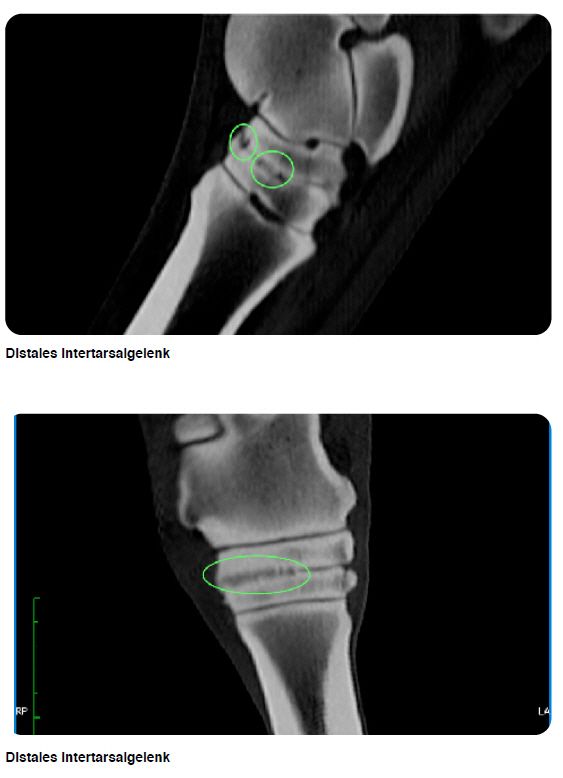
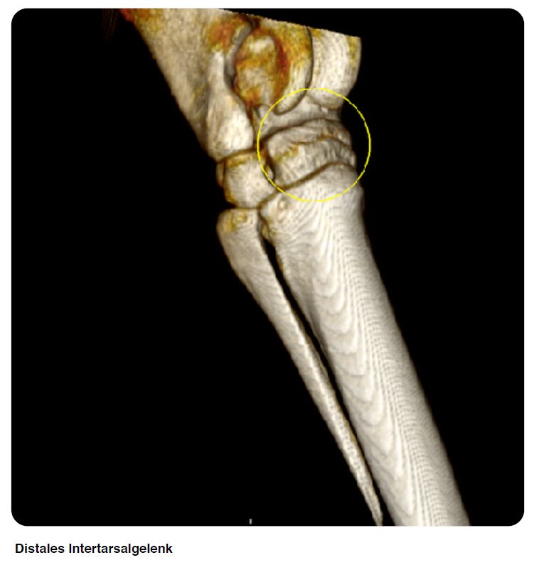
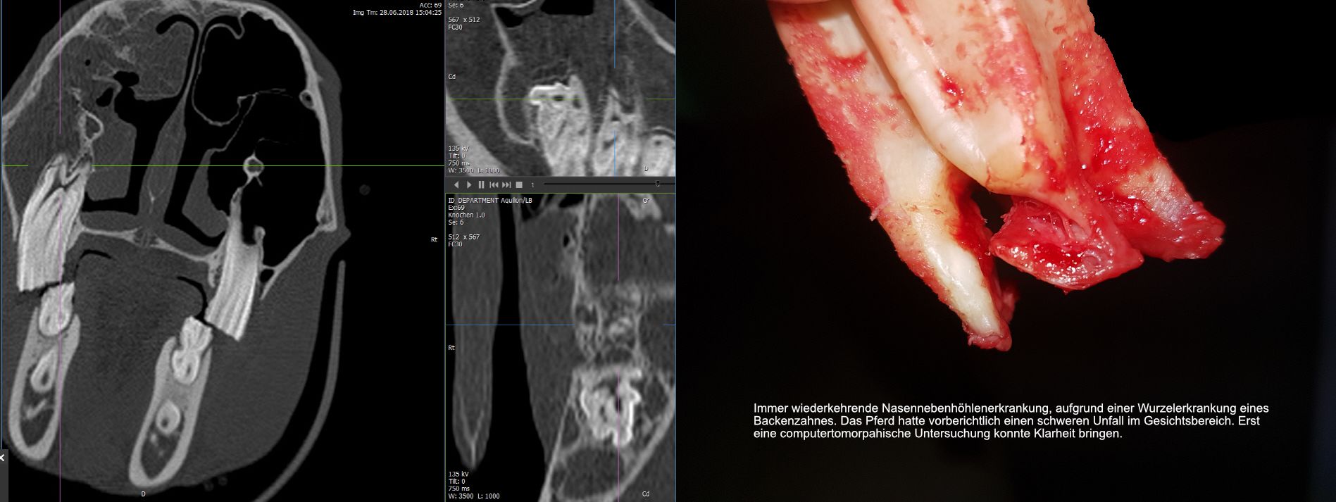
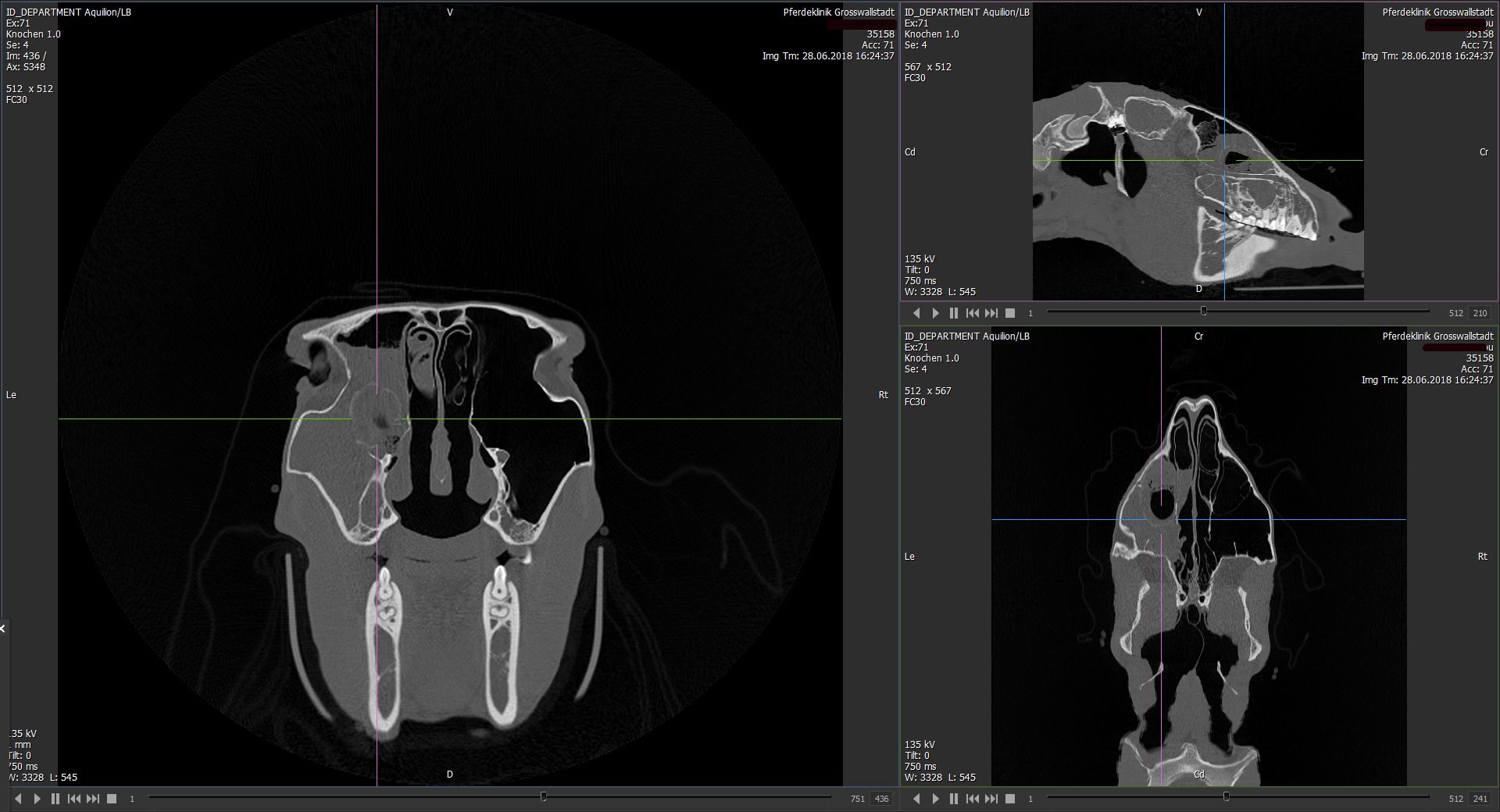
A computed tomography is enormously helpful when it comes to the exact localization of pathological changes and thus to the best planning of the surgical procedure. This is needed, for example, in dental problems, especially involving the paranasal sinuses, or in numerous orthopedic problems such as bone cysts, fissures, and mostly complex fractures. In addition, soft tissues (tendon and ligaments) can also be imaged and highlighted with special contrast techniques. Using contrast techniques it is also possible to image for the first time articular cartilage surfaces and its defects.
This advanced examination method completes our diagnostic range of X-ray, ultrasound, endoscopy, scintigraphy, which, as you know, are always state-of-the-art, to optimally assist you and your horse.
In order to keep the quality of the results at the highest level, a so-called 4-eye principle is routine for us. By teleradiology, the results of the investigation are evaluated not only by our specialist, but also by highly specialized national colleagues.
CT-Screening Video
The Aquillion device from Toshiba has been strengthening the diagnostic capabilities of our clinic for a few weeks now and offers our equine patients an examination at the highest level.
It is a 32-rower, this is considered the "gold standard" in veterinary medicine. Due to its large opening (gantry), the horses’ head and neck can be examined standing, and the distal limbs under general anesthesia, in less than 30 seconds.
Computed tomography (CT) is an multi-dimensional X-ray examination. The generated slice images (0.5 mm thick) provide a detailed 3D view of the affected region of the horse's body.
A computed tomography is enormously helpful when it comes to the exact localization of pathological changes and thus to the best planning of the surgical procedure. This is needed, for example, in dental problems, especially involving the paranasal sinuses, or in numerous orthopedic problems such as bone cysts, fissures, and mostly complex fractures. In addition, soft tissues (tendon and ligaments) can also be imaged and highlighted with special contrast techniques. Using contrast techniques it is also possible to image for the first time articular cartilage surfaces and its defects.
This advanced examination method completes our diagnostic range of X-ray, ultrasound, endoscopy, scintigraphy, which, as you know, are always state-of-the-art, to optimally assist you and your horse.
In order to keep the quality of the results at the highest level, a so-called 4-eye principle is routine for us. By teleradiology, the results of the investigation are evaluated not only by our specialist, but also by highly specialized national colleagues.
APPOINTMENTS FOR CUSTOMERS
Phone
(0) 6022 / 265 970
(0) 6022 / 265 970


")
")